
Trans-esophageal echo-cardiology (TEE) probes. Merely uttering those words can sound intimidating. Even imaging service engineers, who’ve supported diagnostic ultrasound for years, may have never truly felt comfortable handling these devices. Most HTM professionals only handle them when they fail and require service. Let’s talk TEE probe 101, and introduce or reacquaint ourselves with the technology and hardware.
TEE Probe Development
TEE probes are considered a specialty-type of ultrasound probe and their development was out of pure need. A TEE study is performed when a standard, trans-thoracic echo-cardiology (TTE), study is inconclusive. Patients with chronic obstructive pulmonary disease (COPD), obesity, or a condition known as barrel chest have physiology that may limit visibility of cardiac structures using traditional ultrasound techniques. The challenge with ultrasound is that there is a tradeoff between resolution and penetration. To adequately penetrate the thoracic cavity of a patient with barrel chest, resolution can be compromised, thus potentially impacting the accuracy of a diagnosis. There needed to be a method, or tool, developed to obtain high-resolution cardiac images that were not limited by a patient’s physiology. Similar limitations exist within OB-Gyn and urological ultrasound, and are the reasons for which endo-cavity probes were developed. Necessity was, once again, the mother of invention.
Common TEE Probe Models
Although there is no industry standard, model numbers assigned to ultrasound probes typically provide insights into aspects of their design. Consider the following
- L12-3: Linear probe (or array) that has a bandwidth from 3 to 12 Mhz
- C1-6-D: Curved probe (or array) that has a bandwidth from 1 to 6 Mhz (D-style connector)
- 6S-RS: Sector probe (or array) that has a center frequency of 6 Mhz (RS-style connector)
- IC or EV: Inter-cavity or Endo-cavity probe
- X (prefix): Philips X-matrix live 3D volumetric probe (or array)
- Z (Siemens): Live 3D volumetric probe (or array)
- V (GE): Live 3D volumetric probe (or array)
TEE probes are no different. There is usually a “T” in the model number.
| Philips | GE | Siemens |
| X8-2t | 6VT-D | Z6M |
| X7-2t | 6Tc | TE-V5Ms |
| S7-3t | 6T-OR | |
| S8-3t | 9T | |
| S7-2t Omni |
TEE Probe Design
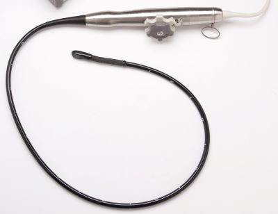
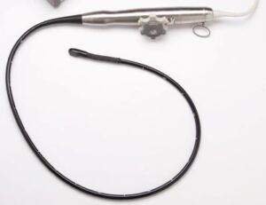
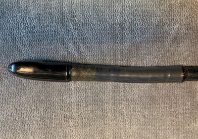
Similar to standard probes, TEE probes have an acoustic lens, acoustic array, scanhead electronics, a wiring harness, connector electronics and a system connector. No different, right? Hardly. All of the components within a standard probe need to be miniaturized and attached to the end of an assembly that can be inserted in the esophagus. The array and scanhead electronics are located in the distal tip at the end of a flexible insertion tube.
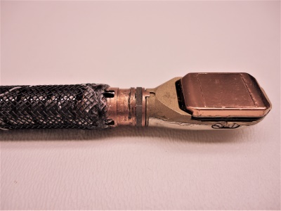
The image to the left shows the acoustic array, ASIC, and electronics contained within the distal tip of a Philips X7-2t TEE probe.
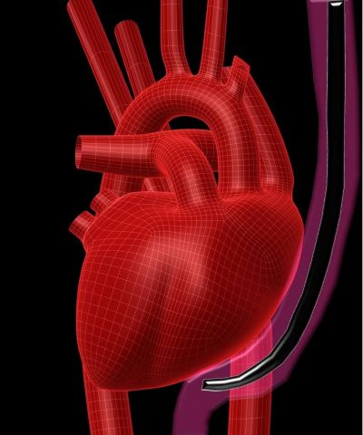
The esophagus is positioned directly behind the heart, which provides an ideal depth for obtaining higher-resolution images. To obtain various cross-sectional images, the distal tip can be flexed in four directions using knobs on the control housing. Buttons on the control housing allow the image orientation to be adjusted (either mechanically on 2D TEEs or electronically on 3D TEEs). The combination of articulation and image orientation allow cardiologists similar freedoms as adjusting the position and angle of a standard trans-thoracic probe.
TEE Probe Wear and Tear
Wear and tear on a TEE probe are much higher than that of any other type of ultrasound probe. The probe is repeatedly exposed to acidic GI fluid for up to eight hours or more. TEE studies can last as little as 30 minutes, for outpatient or bedside studies, or as long as eight hours during a surgical procedure such as a valve replacement. TEE probes must also be high-level disinfected after each use, which is typically performed via immersion in harsh chemical solutions.
Our research shows that after about 100 to 150 uses, the rubber-like materials on a TEE probe, specifically the bending rubber, tend to become more brittle and more apt to perforate if they contact a sharp object. Add to this the fact that the distal tip, bending section and insertion tube may be bitten, dragged across sharp teeth or accidentally damaged during transportation. No bite guard ever created can protect a TEE probe from the gag reflex of a frightened patient who awakens during a TEE study.

Catastrophic TEE Probe Damage
The process of disinfecting a TEE probe, with a break in physical integrity, is one of the most-frequent modes of failure. As such, about 50 to 60% of all TEE probes that arrive at our facility present with gross fluid invasion because of some type of physical damage or improper use. Once fluid (bodily or chemical disinfectant) enters the probe, catastrophic electro-mechanical failures will occur. It only takes a few hours for chemical agents to begin corroding the electro-mechanics within the probe and energizing a fluid invaded probe on a scanner may not just damage the probe, but also induce thousands of dollars of damage to the scanner.
Where TEE Probes are located
TEE probes are typically only in use at larger metropolitan health care facilities with heart failure programs. Smaller metropolitan facilities, or rural and general hospitals, may not have a single TEE probe. For those that do, expect only about 10% of your total probe volume to be TEE probes. That being said, supporting TEE probes may cost as much as supporting the other 90% of your organization’s standard probes.
TEE Probe Support Costs
OEM replacement TEE probes can cost anywhere from $20,000 to $40,000 apiece and you can expect a 70% failure rate per probe per year. There is good news! By using Innovatus Imaging, even catastrophically damaged TEE probes can be restored. There are multiple TEE probe models on which we have 100% repair capabilities and expect repair costs to be only a fraction of the replacement cost (starting at only $500).
Our Approach to Repair
Some repair providers offer to dehydrate fluid invaded probes and take the “dry it and try it” approach. Our goal in addressing fluid invasion is to REPLACE any and all affected components, thus restoring performance. It’s what allows us to offer a six-month warranty on all TEE probes (repaired or exchanged). It’s one of the industry’s longest warranty periods.
How to Reduce Your Support Costs
We’re here to help you NOT NEED our repair services. We’ve also created tools to help you help your teams reduce failure rates and failure severity. Check out our SafeTEE page and learn how adding TEE probes to a preventive maintenance program can make an impact.
Comments 2
Matt great info here. As you know I spent many years dedicated to the Cardiac Ultrasound World. You have provided great info and I would only add a few other thoughts.
1. In the Ultrasound world this the the most expensive transducer there is and it is also the most vulnerable transducer there is to mishandling.
2. One of the most common failures to these probes are bite holes in the endoscope portion of the probe. It is important for the Field Service Engineers to understand this and to try and look for this type of failures.
3. Most OEM’s provide bite guards for this Probe. However, many time’s the bite guard is not utilized.
4. It is important to have an Autopsy of these failed probes. Many times the Customer is at fault for these failures.
5. Once a TEE probe sheathing is compromised and fluid intrusion has happened it is very expensive to repair. This is why preventative measures are so important in TEE Care.
Author
Thanks for taking time to read our post and for sharing your thoughts, Joe. You’ve jumped ahead to our next post, which will present some of the items that you mentioned.