
Is it 3D, 4D, or 3D/4D, and what is 5D Ultrasound?
You may be surprised to know that the first 3D ultrasound image was acquired back in 1986. Saying that 3D ultrasound technology has advanced over the last 30-some years is an understatement. In its infancy, 3D ultrasound images were acquired by sonographers manually moving a standard ultrasound probe across the anatomy, while sensors determined the probe’s relative change in position. Images were only static (single image), and they were not anywhere near real time. Fast-forward to 2010. We’re able to view a beating heart in 3-dimensions, rotate the live image in any direction, and non-invasively assess cardiac functions in real-time using 3D matrix array probes. But how did we get from point-A to point-B, and is that bridge technology still relevant?
Bridge Technology
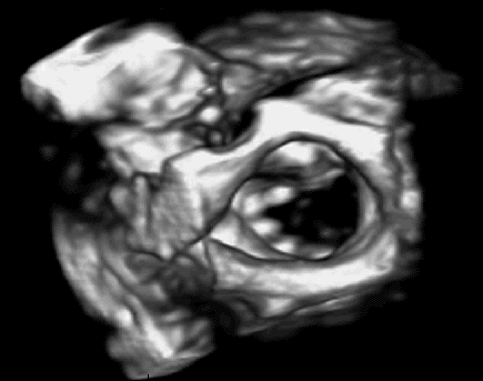
In between the manually acquired 3D images of the 1980s and 90s, and the live-3D images of the 2000s exist mechanically driven 3D probes. Probes, such as the GE RIC5-9-D or Philips 3D9-3v, utilize a traditional 2D array, which is mechanically swept across, or oscillated back and forth, while multiple single images (or slices) are acquired by the scanner. The multiple images are then integrated with one another and assembled to render a 3D image. Volume data acquisition, analysis, and display occur in, almost, real time. The scanner’s software interpolates any gaps in information between each slice to create a smooth image. Just like the images acquired by today’s 3D matrix array probes, mechanically acquired images can be sliced, diced, or rotated in free space.
| OEM | Mechanical 3D Probe Models | Matrix, Live 3D Probe Models |
| Philips | 3D9-3v, 3D6-2, V6-2 | X6-1, X7-2, X3-1, X5-1, X7-2t(TEE) X8-2t(TEE) |
| GE | RIC5-9-D, RAB4-8-D, RAB-6-D | 4V-D, 4Vc-D, 6VT-D(TEE) |
| Samsung/Medison | V5-9, CV1-8A, EV2-12, EV2-10A | |
| Siemens/Acuson | 7CF1, 9VE3, 9VE4, 8VC3 | 4Z1c, Z6M(TEE) |
| Canon/Toshiba | 9CV2 | i6SVX2(Sector), i6SVX2(TEE) |
Despite the popularity of high-tech matrix array probes, mechanically driven probes still maintain a strong market presence. The majority are used in ob-gyn, abdominal, and small parts applications. In fact, the design of the mechanically driven probes of the late 1990s and early 2000s isn’t much different than current models. One of the reasons for their continued popularity is cost per benefit. The applications, in which mechanical probes are used, do not merit the added costs associated with the technology needed to support matrix array probes.
One of the reasons for developing matrix array technology was visualization of cardiac activity. Based on the amount of and the speed at which cardiac activity occurs, a mechanically driven probe would be unable to effectively capture cardiac volumetric data. In the time that a mechanical probe would perform one acquisitional sweep, the activity level within the heart would have changed considerably. Activity in the abdomen, small parts, and womb occur at a very slow pace when compared to the heart. Existing, cost-effective technology is more than able to provide radiologists with the image quality and volumetric data that is needed to make accurate diagnoses (and parents with a 3D image of baby).
Another reason for their continued popularity is related to physical size. The most popular models of mechanical probes are used in intercavity studies. Current matrix array technology is quite sophisticated, and it does increase probe size when compared to 2D counterparts (ex. Philips X5-1 versus S5-1). Rather than the 80 acoustic elements in an S5-1, the X5-1 has over 3000. Applying the same technology to an intercavity probe would make an already cumbersome probe that much more.
Mechanical probes will remain in the marketplace until both the cost and physical size of matrix array probes and technology decrease. We’ve already seen new matrix array probes designed for abdominal and small parts use, but the cost of both the probes and the scanner technology makes it challenging for departments to justify the added costs. There is minimal increased reimbursement for a 3D ultrasound versus a traditional 2D ultrasound study based on minimal added “clinical” benefit.
So, what exactly is 3D, 4D, 3D/4D, and now 5D ultrasound?
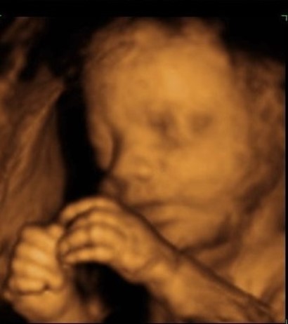
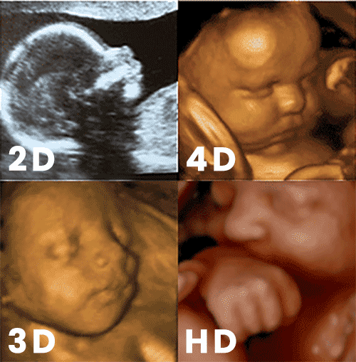
In general, its all the same, just with minor tweaks to the details. 3D ultrasound refers to a single static 3D ultrasound image. 4D ultrasound is the addition of time. Consider it live, or semi-real time, acquisition and display of 3D ultrasound data that allows for volume imaging and volume calculation. 3D/4D is a general term that refers to ultrasound probes that can be used to acquire both a 3D image and a live 3D (or 4D) volumetric image. 5D ultrasound is the latest buzzword. Also known as HD ultrasound, 5D uses software that makes images appear smoother, as if captured using higher resolution, and also gives the image a flesh-colored tone. 5D seems to be targeted at the Obstetrics market. The purpose seems to be purely marketing in that the image of the in-utero baby appears more natural and more photogenic.
Think of it this way.
- With 2D ultrasound, the length and width of an artery can be visualized
- Using 3D ultrasound, it is possible to visualize depth to the artery
- 4D ultrasound enables live visualization of the artery and enables users to calculate the volume within the artery
- 5D/HD just makes the artery look “pretty” or flesh-colored
Relative to 4D and 5D, there are no changes in probe or scanner technology that actually enables a 4th or 5th dimension of imaging.
How can we help?
Innovatus Imaging supports your most popular 3D imaging probe models, whether mechanical or matrix technology. Multiple models are fully repairable, meaning that you’re not presented with costly options for replacement…Just cost-effective repair solutions. All 3D probes are backed by a 6-month warranty… One of the longest in the industry. For more information about probe repair and our capabilities, please reach out to our Customer Care team or visit www.innovatusimaging.com/ultrasound
Comments 2
We’d like to find out about your 4-D ultrasounds.
Author
Happy to share our capabilities and approach to repair. I will reach out to the email address that you provided.